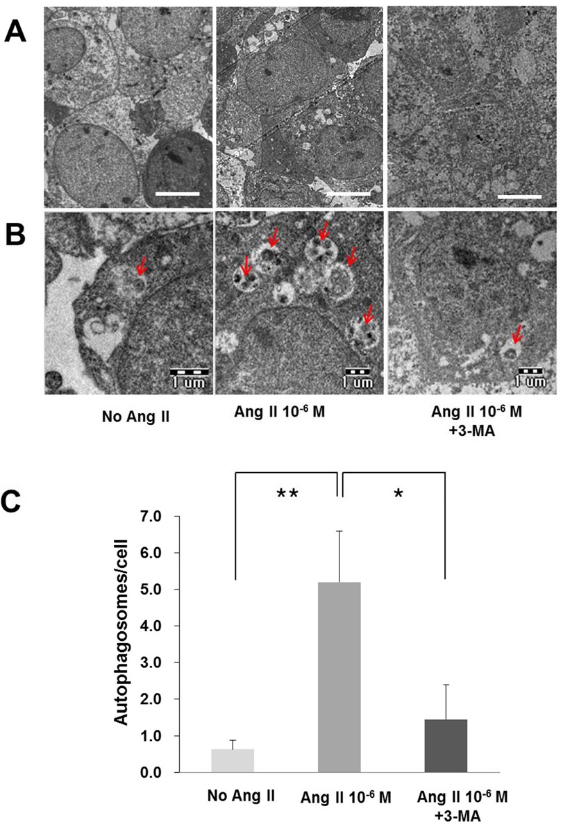
Fig. 4. Effects of Ang II and 3-methyladenine (3-MA) on autophagosomes formation in podocytes detected using transmission electron microscopy. Equal numbers of mouse podocytes were incubated. More autophagosomes were observed in Ang II-treated cells (A, magnification, ×3000; Bar = 10 μm). The numbers of autophagosomes (arrows) are increased with Ang II treatment; 3-MA (2 mM) attenuates autophagosomes formation at 12 hours (B, magnification, ×10000; Bar = 1 μm). The numbers of autophagosomes are counted and quantified in 10 random podocytes (C). Data on the analysis of autophagosomes ratio of podocytes are presented as mean ± SD (n = 5). Control (100%); compared to conditions without Ang II. *P<0.05 and **P<0.01 versus control.